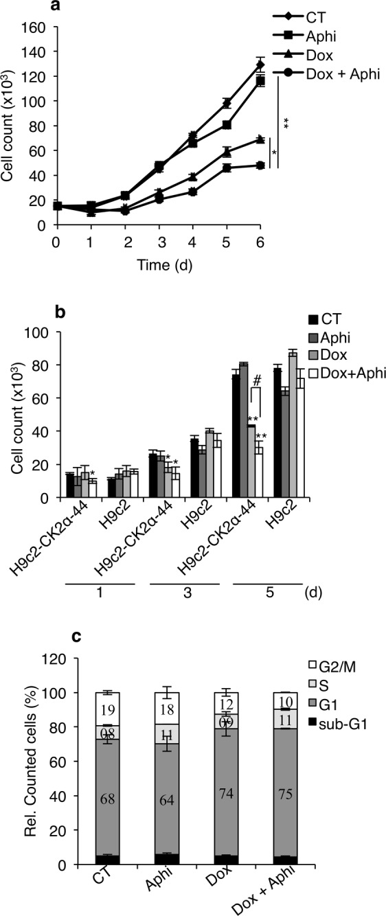
Figure 3.

Analysis of cell proliferation following induction of mild DNA replication stress. (a) H9c2-CK2α-44 incubated with 1 µg/ml doxycycline for three days were subsequently re-seeded and treated with 0.1 µM aphidicolin (Aphi) for increasing amounts of time as indicated in the figure. Control experiments (CT) refer to cells grown in the presence of 0.1% DMSO for up to five days. Cell proliferation was determined by hemocytometer counting. Experiments were carried out three times in triplicates obtaining similar results. Results of one representative experiment are shown +/− STDEV, *P < 0.05, **P < 0.005. (b) Comparison between H9c2-CK2α-44 and H9c-2 cells with respect to proliferation efficiency. Cells were treated essentially as described in (a) for the indicated times. Mean values+/− STDEV of one representative experiment out of three is shown. *P < 0.05, **P < 0.0005 with respect to CT, #P < 0.01. (c) H9c2-CK2α-44 cells left untreated or treated with 1 µg/ml doxycycline for three days were co-treated with 0.1% DMSO or 0.1 µM aphidicolin for additional five days. Cells were analyzed by flow cytometry following propidium iodide staining (PI) and events were quantified and expressed in percentage.
