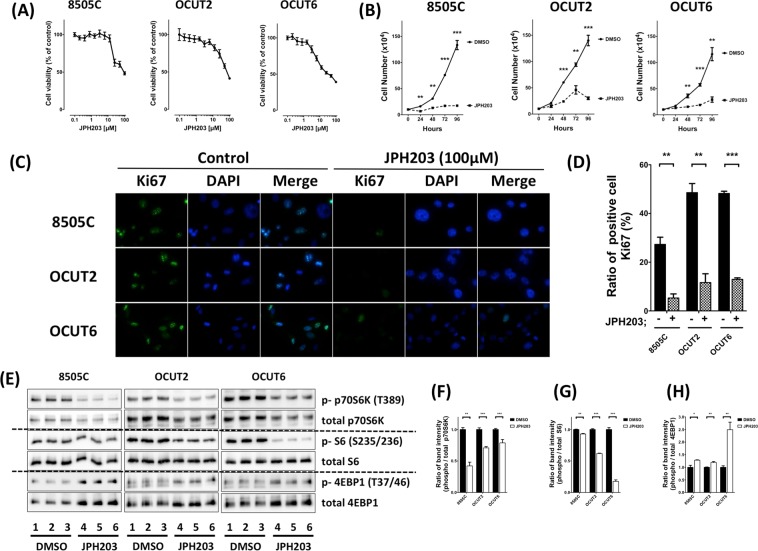
Figure 3.
Effect of JPH203 on cell viability (A) and tumor cell growth (B) in 8505C, OCUT-2 and OCUT-6 cells. (B) Three cells were treated with vehicle (DMSO) or JPH203 at increasing doses of 100 µM for 24, 48, 72, or 96 hours. Cells were harvested, and cell numbers were counted. (C) Detection of Ki67 (green) by immunofluorescence analyses in 8505C, OCUT-2, and OCUT-6 cells. DAPI nuclear staining was shown in panel (blue). Ki67 and DAPI were merged and shown in right panels. (D) Quantification of Ki67-positive cells. After treatment with 100 µM JPH203 for 24 h, 8505C, OCUT-2, and OCUT-6 cells had significantly lower numbers of cells stained for Ki67 than those treated with DMSO as control. (E) Western blot showing phosphorylation of p70S6K, S6, and 4EBP1 in 8505C, OCUT-2, or OCUT-6 cells with 100 µM JPH203 for 24 h or DMSO as control (F–H). The band intensities were quantified and compared in these mTOR signals (*p < 0.05, **p < 0.01, ***p < 0.001).

