Fig. 6.
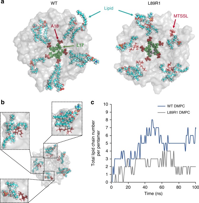
L89R1 side chain steric clashes restrict lipid-chain penetration within the NP. a Representative MD simulation top pore view snapshots of WT and L89R1 (transparent gray surface view), residues L17 (green spheres, pore constriction site), and A18 (red spheres, NP bottom) are highlighted. NP-associated lipids are shown in cyan sticks. The bulk of the bilayer lipids is hidden for clarity. All five lipid chains make direct contact with A18 in the WT channel and only a couple of them with the same residue in L89R1 channel as MTSSL (red sticks) clashes with lipids obstructing their entrance to the NPs. b Close-up views (dashed line boxes) of some different steric-clash interactions between MTSSL and lipids on L89R1 TbMscL. c Comparison of the total number of lipid chains residing within the NPs (per single TbMscL pentamer) over time (100 ns atomistic MD simulation), between WT (blue line) and L89R1 (gray line) TbMscL channels

