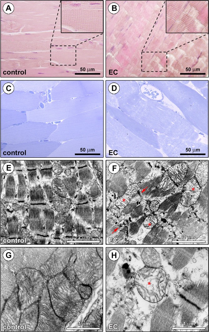
Figure 1.
Morphological evaluation of ex vivo eccentric contraction (EC)-induced skeletal muscle damage model. (A,B) Hematoxylin and eosin staining testifying the normal structure of control muscles and the presence of tissue damages in EC-injured muscles. Higher magnifications of the boxed areas are shown in the insets. Note Z-disc smearing/streaming and focal loss of myofilaments in EC muscles. (C,D) Semithin sections of control and EC-injured muscle samples stained with toluidine blue and observed by light microscopy confirming the substantial EC-induced structural abnormalities. (D) Swollen and round-shaped myofibers and intramyofiber and interstitial edema are evident in EC-injured samples. (E-H) Ultrathin sections of control and EC-damaged muscle samples stained with UranyLess and bismuth subnitrate solutions and examined by TEM. (F,H) EC-injured myofibers exhibit an evident disorganization of sarcomere structures and Z-disc disruption (arrows), as well as swollen mitochondria with disarranged or even missing cristae (asterisks). Images are representative of at least 6 sections from each of 5 control and 5 EC-injured muscles.

