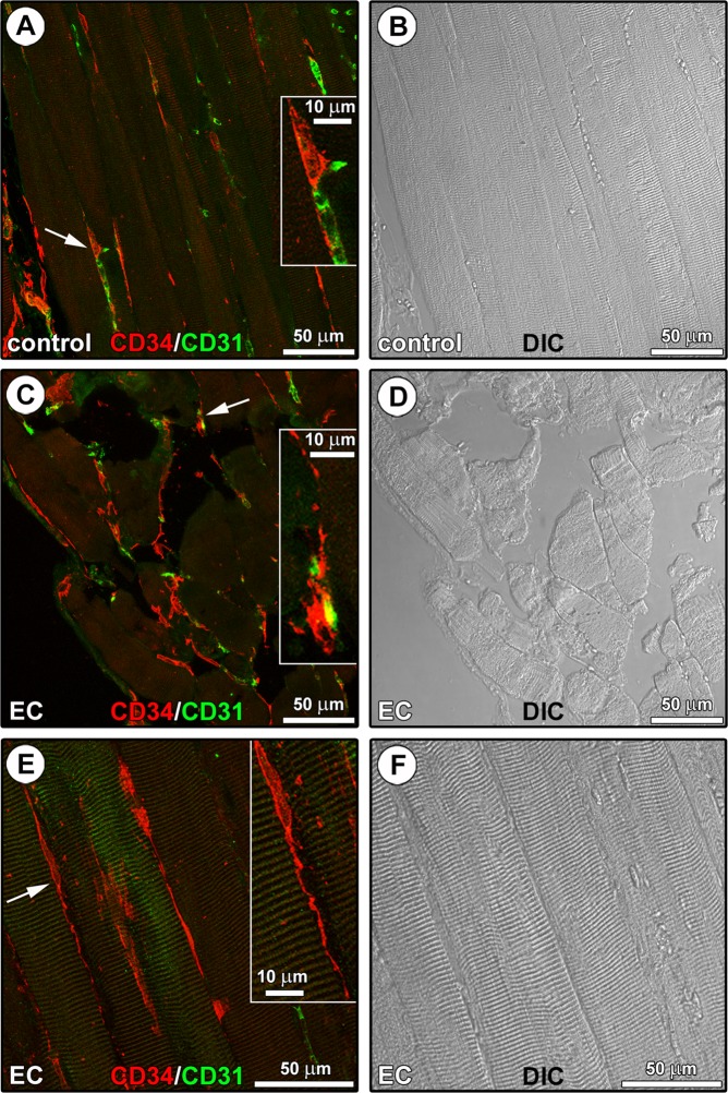
Figure 3.
CD34/CD31 confocal immunofluorescence staining of paraffin-embedded tissue sections from control and eccentric contraction (EC)-damaged skeletal muscles. (A–F) Representative fluorescence images (A,C,E) and differential interference contrast (DIC) images (B,D,F) acquired simultaneously to allow a better appreciation of the tissue structure. (A) In control muscles, CD34+CD31− telocytes (TCs) with distinctive long and thin varicose cytoplasmic prolongations (telopodes) are located in the interstitium alongside the myofibers and in the close vicinity of CD31+ vascular structures (arrow and higher magnification in the inset). (C,E) An extensive CD34+CD31− TC meshwork is evident among myofibers in EC-damaged muscles. Insets: magnifications of the areas indicated by arrows; note the close vicinity of a CD34+CD31− TC to a CD34+CD31+ blood capillary vessel (C), and a CD34+CD31− spindle-shaped TC sending a distinctive moniliform telopode over long distance in the interstitial space among two myofibers (E). Images are representative of at least 6 sections from each of 5 control and 5 EC-injured muscles.

