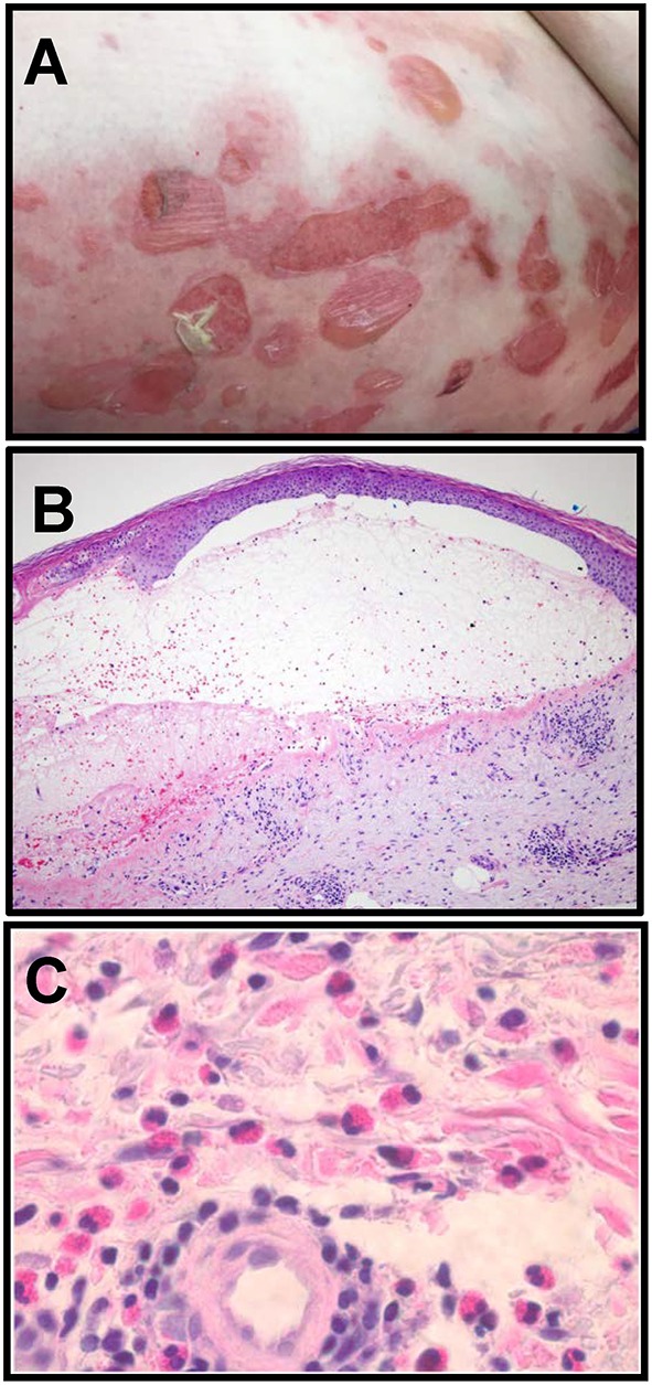
Figure 1.

Clinical and histologic characteristics of Bullous pemphigoid. (A) Clinical presentation of BP with tense, fluid filled blisters occurring on areas of erythema and normal skin, frequently associated with urticarial plaques. (B) Blisters correspond histologically to a subepidermal separation at the basement membrane zone (BMZ) with eosinophils observed in the superficial dermis and the blister cavity. H and E, 100x. (C) Eosinophils in the deep perivascular infiltrate in a lesional biopsy from a BP patient. H and E, 400x (Images in B, C are courtesy of Dr. Brian L. Swick, University of Iowa).
