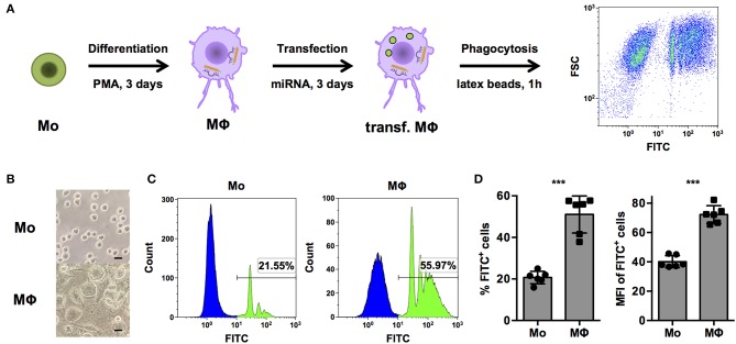
Figure 1.
PMA-activated THP-1 cells allow investigation of phagocytosis of opsonized latex beads by human MΦ. (A) Schematic representation of the workflow for differentiation, transfection, and analysis of phagocytosis of FITC-labeled, opsonized latex beads for high-content screening of a miRNA library and subsequent experiments. (B–D) Bright-field microscopy and FACS analysis of untreated (Mo) or PMA-activated (MΦ) THP-1 cells. (B) Microscopic images were acquired with an AxioObserver Z1 microscope (Zeiss) using a 40× objective. Scale bars represent 20 μm. (C) Histogram plots of one representative experiment and (D) quantification of the percentage of FITC+ (left) and mean fluorescence intensity (MFI) of FITC+ cells (right) of n = 6 independent experiments. In (D), values from each experiment are indicated by symbols and the mean ± standard deviation (SD) are indicated by a bar and whiskers. Statistical analysis was performed using Student's t-test (***p < 0.001).

