Figure 2.
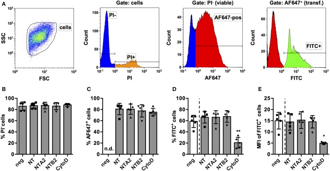
Establishment of a FACS-based assay to screen for miRNAs for effect on phagocytosis of PMA-activated THP-1 MΦ. (A) Activated THP-1 Mq were identified by forward scatter (FSC) and side scatter (SSC), and viable cells were selected by excluding PI+ cells. Viable cells were analyzed for successful transfection by positive AF647 staining of the control NT siRNA. Within the PI− AF647+ population, phagocytic cells were identified using the FITC signal of fluorescent latex beads. Viability of cells (% of PI− cells; B), transfection efficiency (C), percentage of phagocytic cells (D), and phagocytic activity (MFI of FITC+ cells; E) were measured in cells transfected with the NT siRNA alone or in combination with two non-targeting miRNAs (NTA2 and NTB2) or with cytochalasin D treatment (CytoD). In (B–E), individual values of n = 5 independent experiments are indicated by symbols and the mean ± SD are indicated by a bar and whiskers. Statistical analysis was performed using repeated measures, one-way ANOVA. Dunnett's post analysis was used to calculate p-values adjusted for multiple comparisons with NT-transfected cells as control condition (*p < 0.05; **p < 0.01). In (B), negative control cells (neg) were set as control condition to check if any of the treatments had an effect on viability.

