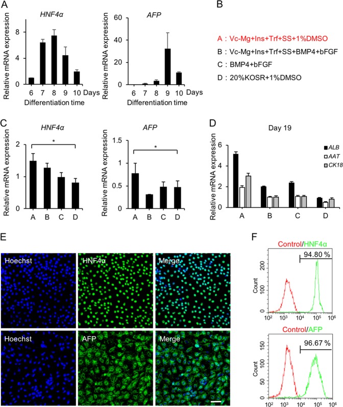
Fig. 3. Differentiation of hESCs into hepatoblasts in defined xeno-free conditions.
a The relative hepatoblast gene (HNF4α and AFP) expression levels of cells differentiated with a previously reported protocol were determined by qPCR at days 6, 7, 8, 9, and 10. b Four protocols were used to induce PE to hepatoblasts. Trf (transferrin, 5 mg/mL); Vc-Mg (L-ascorbic acid 2-phosphate sesquimagnesium salt hydrate, 50 µg/L); Ins (insulin, 10 µg/mL); SS (sodium selenite, 0.1 ng/mL); and BMP4 (10 ng/mL); bFGF (10 ng/mL). c The relative hepatoblast gene (HNF4α and AFP) expression of day 9 differentiated cells in basal medium (DMEM/F12, 1 × L-GlutaMAX, and 1 × NEAA) with different treatments (groups A, B, C, and D) were determined by qPCR. d The relative hepatocyte gene (ALB, AAT, and CK18) expression of day 19 differentiated cells in basal medium (DMEM/F12, 1 × L-GlutaMAX, and 1 × NEAA) with different treatments (groups A, B, C, and D) were determined by qPCR. e Immunofluorescence analysis of HNF4α and AFP expression in group A-induced differentiated cells on day 9. f The expression levels of HNF4α and AFP in group A-induced differentiated cells were determined by flow cytometry on day 9. Isotype control antibodies were used as controls. *p < 0.05, **p < 0.01; data are represented as the mean ± SD. Scale bar, 100 µm.

