Figure 4.
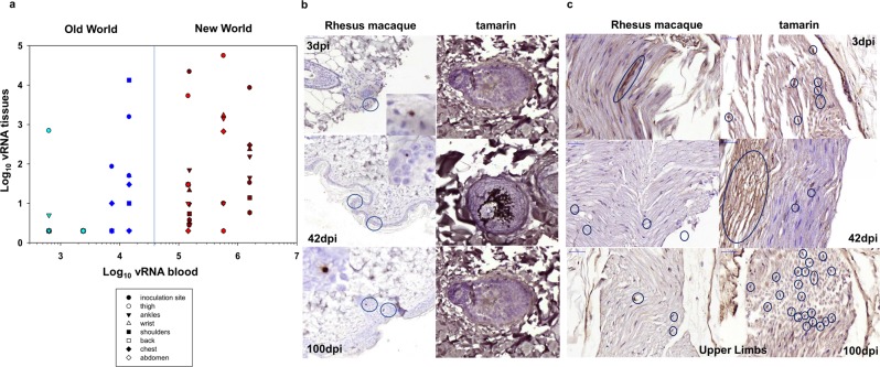
(a) Relationship between tissue vRNA and blood vRNA levels in individual monkeys in multiple skin tissues 3 dpi. Symbols representing each tissue sampled are shown in the key. Colour code: marmosets (dark red), tamarins (red), Indian rhesus macaque (blue) and Mauritian cynomolgus macaque (light blue). (b) Localisation of ZIKV in skin biopsies. Representative images of RNAscope ISH detection of ZIKV RNA within FFPE skin punch biopsies for rhesus macaque and red-bellied tamarins. Sections were collected from each species from defined sites across the body post-mortem 3, 42 and 100 dpi. Images shown are from the inoculation site on the back of the neck (tamarin P1; 42 dpi and tamarin P8, 100 dpi) and ankle (tamarin P5, 3 dpi). Image (x20 magnification), brown stained ZIKV RNA positive cells identified within both epidermal and dermal layers of the skin but primarily in the external root sheath of hair follicles. Numerous ZIKV RNA stained cells with a dendritic cell morphology were present within red-bellied tamarins 100 dpi. (c) Immunohistochemical staining for NS-1 protein in peripheral nerves. All images x40 magnification shown for section across areas of upper limb in rhesus macaques (left panels) and tamarins (right panels) at 3, 42 and 100 dpi. Clusters of nerve fibre cells are visible 3 dpi.

