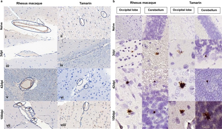
Figure 5.
Impact of ZIKV in brain tissue. (a) Immunohistochemical staining for the tight junction protein zonula occludens-1 (ZO-1). Characteristic staining within the walls of blood vessels present in white and grey matter of Old and New World primate cerebral cortex. Within the blood vessels of naïve animals, rhesus macaque and tamarin ZO-1 is detected throughout the vessel walls (i,ii). 3 days post infection (iii,iv) levels of ZO-1 staining are greatly reduced in both species. By 42 (v,vi) and 100 (vii,viii) days post-infection (dpi) ZO-1 staining is present within blood vessel walls but staining exhibits a fragmented pattern in contrast to continuous staining in naïve animals. (b) RNAscope detection of ZIKV RNA in CNS. Representative images within FFPE sections of central nervous system (CNS) of occipital lobe and cerebellum collected post-mortem from rhesus macaques or red bellied tamarins 3, 42 or 100 days post-infection (dpi). Comparable distribution of ZIKV RNA positive cells (brown stain, x20 magnification) within the CNS of both Old and New world species. Viral foci detected as early as 3dpi which remained detectable through to 100 dpi. ZIKV positive cells identified within both white and grey matter with a predominantly glial cell morphology associated with neuronal cell bodies.

