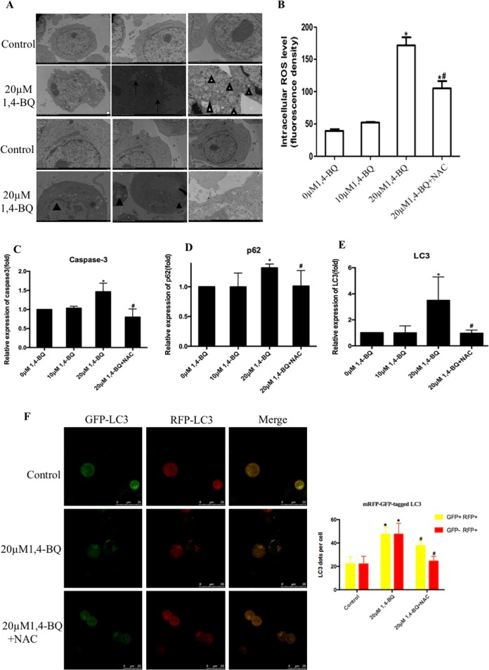
Fig. 4. 1, 4-BQ induced autophagy and apoptosis via activating oxidative stress.
a The images of TEM showed that 1, 4-BQ induced autophagy and autophagosome accumulation. White arrow, double-membrane autophagosome (White triangle) and single-membrane autolysosome (Arrows). TEM ultrastructural analysis showed that 1, 4-BQ activated apoptosis and the formation of apoptotic body. The chromatin margination, nuclear fragmentation and apoptotic body were observed (Black triangle). b The level of oxidative stress was measured by fluorescent microplate reader. c–e The toxic effect of 1, 4-BQ on the expression of autophagy- and apoptosis-associated genes was investigated by qRT-PCR. f After treating 1, 4-BQ and oxidative stress inhibitor (NAC), autophagy evaluated by mRFP-GFP-LC3 virus

