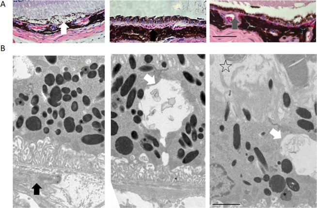
Figure 2.
Nrf2−/− mice can develop atrophic AMD-like phenotypes after mild light exposure. (A) The H&E histology showed the dry AMD phenotypes on light exposure induced Nrf2−/− model. From left to right: vacuole, drusen-like deposit, and choroid vascular changes, Scale Bar 50 µm. (B) The TEM images showed the dry AMD phenotypes on light damaged Nrf2−/− model. (White Arrow: vacuole, black arrow: drusen-like deposit, asterisk: lipofuscin granule, and star-shape: PRC cells detachment). Scale bar: 2 µm. Original images were presented in Supplementary Fig. S6.

