Figure 2.
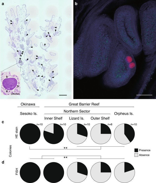
Appearance and occurrence of CAMAs in the coral Acropora hyacinthus. (a) Numerous CAMAs (indicated by arrows) are visible in a histological section stained by hematoxylin and eosin of a colony from Sesoko, Japan. In the panel, small photo shows close-up of a CAMA located in a mesentery of a polyp. (b) Right panel shows two of CAMAs (red) distributed with Symbiodiniaceae (green) in the tentacle (coral tissue: blue) of a sample from the Inner Shelf, GBR using FISH. (c,d) Pie diagrams showing the proportion of colonies sampled that contained CAMAs at five sites in Japan and Australia based on the detections of (c) HE stain and (d) FISH. (c) A significantly higher proportion of Sesoko Is. samples contained CAMAs (including basophilic and eosinophilic) than samples collected from other sites (Lizard Is., Outer Shelf northern GBR site, and Orpheus Is). (d) In the FISH experiment, both proportions of CAMA in samples from Sesoko Is. and Inner Shelf were also significantly higher than in samples from other three sites (**p < 0001; Fisher’s exact test followed by a Benjamini–Hochberg false discovery rate correction). Scale bars indicate 600 µm (a), 50 µm (small panel in a) and 100 µm (b).

