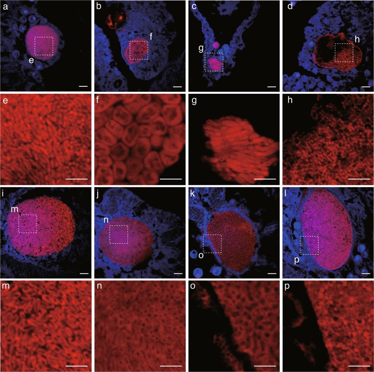
Figure 5.
Morphological variation of bacteria housed within different CAMAs. Note that bacteria within each CAMA are morphologically similar. Dotted lines (in a–d,i–l) delineate regions magnified in close-up images (e–h,m–p). Overall, five bacterial morphologies were detected: rod-like (a,e), pleomorphic (b,f), long rods (c,g), filamentous-like (d,h), rod-shaped with spore-like structures (i,j,m,n), and putative amorphous masses (k,l,o,p). Scale bars indicate 10 µm (a–d,i–l) and 5 µm (e–h,m–p).

