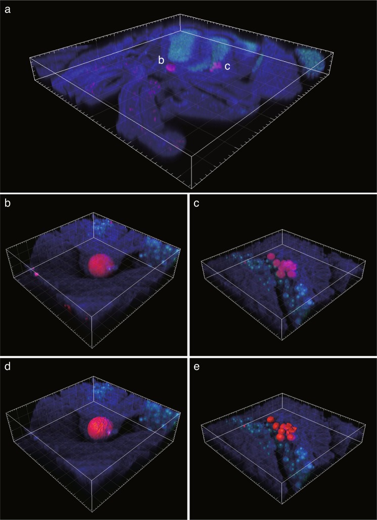
Figure 6.
Three-dimensional (3D) images of CAMAs (red) within a tentacle of the coral Acropora hyacinthus as visualized using FISH. (a) Section of tentacle showing localization of two types of CAMAs (10x magnification). (b) Single aggregation of bacteria in a large structure within the epithelium (40x magnification; composed of 92 z-stack images). (c) Multiple aggregations of bacteria in smaller structures within the gastrodermis (40x magnification; composed of 56 z-stack images). 3D rendering of CAMAs (d,e) reconstructed from 3D images in (b,c). Coral tissue and Symbiodiniaceae appear as blue and green structures, respectively.

