Figure 4.
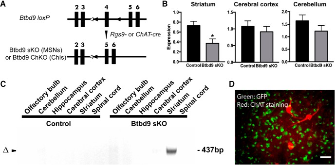
Generation of conditional KO mice and validation of the loss of Btbd9 in the striatum by qRT-PCR. A, Schematic diagram of the generation of conditional KO mice. Filled boxes represent exons. Filled triangles indicate loxP sites (around the 4th exon of the Btbd9 gene). Open triangles indicate the FRT sites that were incorporated to remove the neo cassette. Btbd9 loxP mice were crossed with Rgs9-cre or ChAT-cre mice to obtain double heterozygotes. The double heterozygotes were crossed with Btbd9 loxP homozygotes to obtain Btbd9 sKO or Btbd9 ChKO mice. In conditional KO mice, exons 4 is deleted in specific types of neurons where cre is expressed. Recombination occurs in these cells, while other brain regions and the rest of the body still retain the intact exons. B, Btbd9 sKO mice showed a decreased level of Btbd9 mRNA in the striatum, but not in the cerebral cortex and cerebellum. Bars represent means plus SEs; *p < 0.05. C, Tissue-specific deletion of Btbd9 exon 4 in Btbd9 sKO mice was confirmed by PCR using DNA isolated from each brain region. The deletion was detected only in the striatum of Btbd9 sKO mouse as predicted (Δ). D, A representative immunohistochemical image of a coronal section of the striatum from an Rgs9-cre/GFP mouse. Scale bars represent 25 µm. Enlarged images captured with a 40× objective lens showed that the ChAT staining (red) did not overlap with GFP staining (green). The results suggest that Rgs9-cre does not have cre-mediated recombination in ChIs.

