. 2019 Jul 23;5(3):186–204. doi: 10.3934/microbiol.2019.3.186

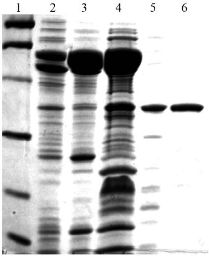
Figure 2. SDS-PAGE gel of RFAP synthase purification. Protein samples were boiled in the presence of 7.5% 2-mercaptoethanol in SDS-PAGE sample buffer and loaded onto a 12% polyacrylamide gel. The gel was stained with Coomassie Brilliant Blue R-250 (Bio-Rad). Lane 1, Molecular mass markers. Lane 2, CFE (8 µg). Lane 3, heated CFE (10.2 µg). Lane 4, hydroxyapatite fraction (17 µg). Lane 5, Mono Q fraction (2 µg). Lane 6, Superdex 75 fraction (2 µg). The bands in lane 1 indicate the positions of the following molecular mass markers: phosphorylase b (97.4 kDa), bovine serum albumin (66.2 kDa), ovalbumin (45 kDa), carbonic anhydrase (31 kDa), soybean trypsin inhibitor (21.5 kDa), and lysozyme (14.4 kDa).

Figure 2.