. 2019 Jul 23;5(3):186–204. doi: 10.3934/microbiol.2019.3.186

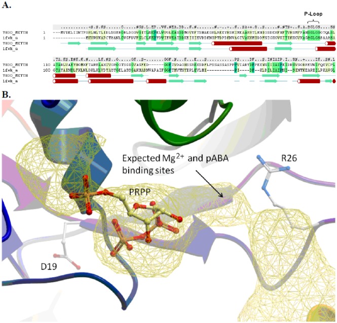
Figure 5. Computational molecular model of MTH0830 RFAP synthase. A: Secondary structure and sequence alignment of the N-terminus of MTH0830 (Y830_METTH) (from residue 1 to 215) and homoserine kinase template structure (pdb code: 1fwk) [44]. The secondary structure elements are colored green (beta Sheet) and red (alpha helix). The secondary structure of MTH0830 was calculated using the DSSP method [45], and the secondary structure of 1fwk is shown as defined in the Protein Data Bank file. B: The predicted PRPP and pABA binding pocket. The MTH0830 model is displayed in ribbon colored from the N to C terminus (blue – red), residues D19 and R26 are displayed as a stick representation. The docked PRPP ligand is displayed in stick representation with carbon atoms colored yellow. The binding pocket is displayed as a yellow mesh, and the predicted sites for pABA and Mg2+ are annotated.

Figure 5.