Fig. 1.

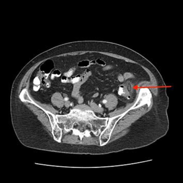
A 20 × 10 × 10 mm fat-density ovoid lesion with a hyperattenuated center (central dot sign) abutting the anterior wall of the proximal sigmoid colon (red arrow) is seen on the CT scan of the abdomen and pelvis.

A 20 × 10 × 10 mm fat-density ovoid lesion with a hyperattenuated center (central dot sign) abutting the anterior wall of the proximal sigmoid colon (red arrow) is seen on the CT scan of the abdomen and pelvis.