
Figure 4.
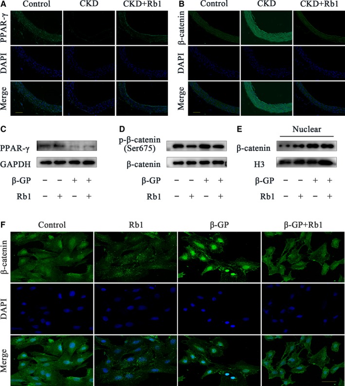
Rb1 activates PPAR‐γ and down‐regulates the Wnt/β‐catenin pathway. A and B, Representative immunofluorescence staining of PPAR‐γ and β‐catenin in rat aortas (scale bar: 100 μm). C‐E, Representative Western blot bands of PPAR‐γ (C), p‐β‐catenin/ β‐catenin (D), nuclear β‐catenin (E) with or without 40 μmol/L Rb1 in the presence or absence of β‐GP for 6 h. F, Confocal microscopy of the immunofluorescence staining of β‐catenin in VSMCs with or without 40 μmol/L Rb1 in the presence or absence of β‐GP for 6 h (scale bar: 100 μm). CKD indicates chronic kidney disease; DAPI, 40,6‐diamidino‐2‐phenylindole; β‐GP, β‐glycerophosphate; H3, histone‐H3; GAPDH, glyceraldehyde‐3‐phosphate dehydrogenase