
Figure 6.
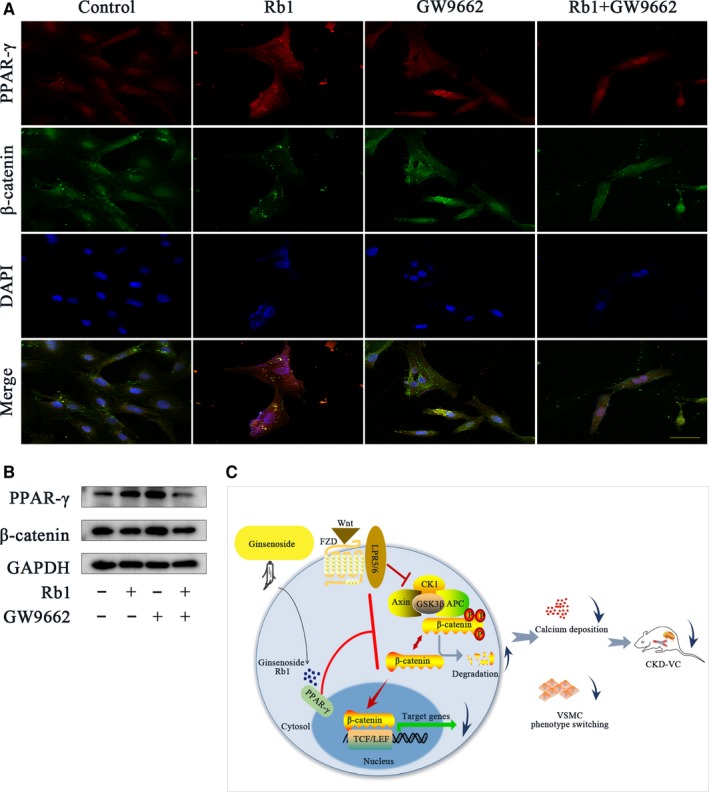
Rb1 inhibits the Wnt/β‐catenin pathway through the activation of nuclear receptor PPAR‐γ. A, Confocal microscopy of the immunofluorescence staining of PPAR‐γ (red) and β‐catenin (green) with different treatments for 24 h: 5 μmol/L GW9662 and 40 μmol/L Rb1 (scale bar: 100 μm). B, Representative Western blot bands of PPAR‐γ and β‐catenin in VSMCs treated for 24 h: 5 μmol/L GW9662 and 40 μmol/L Rb1. C, Schematic diagram of the effect of Rb1 on VSMC calcification. DAPI indicates 40,6‐diamidino‐2‐phenylindole; GAPDH, glyceraldehyde‐3‐phosphate dehydrogenase