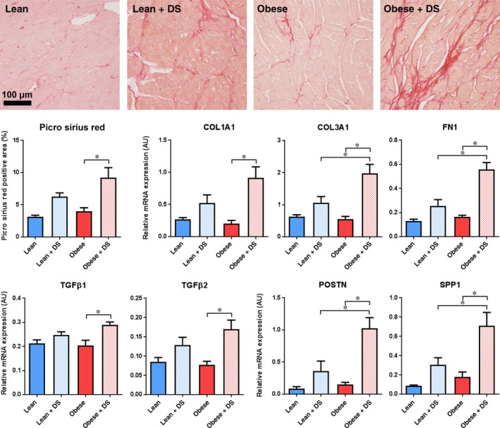
Figure 6.

Fibrosis and TGFβ signalling. (A) Typical examples and (B) quantification of histological staining for cardiac collagen deposition (Picro Sirius Red). n = 6‐7, * P < .05. (C) QPCR analysis of cardiac transcription of extracellular matrix components COL1A1, COL3A1 and FN1, (D) activators of TGFβ signalling TGFβ1 and TGFβ2 and (E) transcriptional targets of TGFβ signalling POSTN and SPP1. n = 5‐7, * P < .05
