
Figure 1.
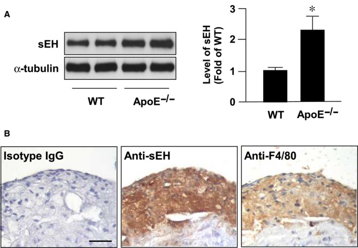
sEH expression is increased in atherosclerotic lesions. Aortas were collected from 5‐ month‐old wild type (WT) mice and apolipoprotein E knockout (Apoe‐/‐) mice. (A) Western blot analysis of sEH and a‐tubulin. Data are mean ± SD from 5 mice. *P < 0.05 vs. WT mice. (B) Immunostaining of macrophage foam cells with (left) control normal rabbit IgG, (center) anti‐sEH, and (right) anti‐F4/80 antibody. Cell nuclei were stained with hematoxylin. Bar = 50 μm