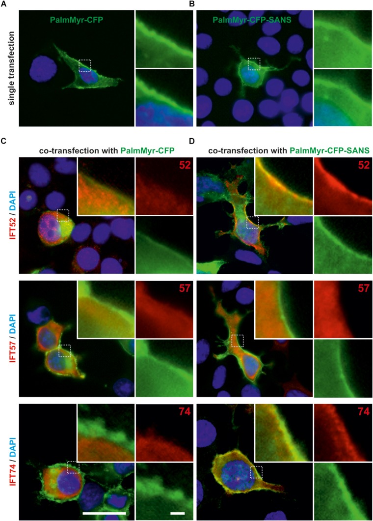
FIGURE 3.
Membrane targeting assay in HEK293T cells revealed in vivo translocation of IFT52 and IFT74 by PalmMyr-CFP-SANS. (A,B) Fluorescence microscopic analysis of HEK293T cells, singly transfected with MyrPalm-CFP or MyrPalm-CFP. MyrPalm-CFP (A) and MyrPalm-CFP-SANS (both in green) (B), respectively, accumulate at the plasma membrane of single-transfected HEK293T cells. (C,D) Fluorescence microscopic analysis of HEK293T cells, co-transfected with FLAG-tagged IFT20, IFT52, IFT57 or 74, and with MyrPalm-CFP (C) or MyrPalm-CFP-SANS (D), respectively. In the control, no co-localization of FLAG-IFT proteins with MyrPalm-CFP alone was observed. In contrast, FLAG-IFT52 and FLAG-IFT74 co-localized with MyrPalm-CFP-SANS at the plasma membrane, whereas FLAG-IFT20 and FLAG-IFT57 did not. Blue, DAPI staining of nuclear DNA. Images are representative for co-transfected cells from two independent experiments. Scale bars: 25 μm; 2.5 μm in zoomed squares.

