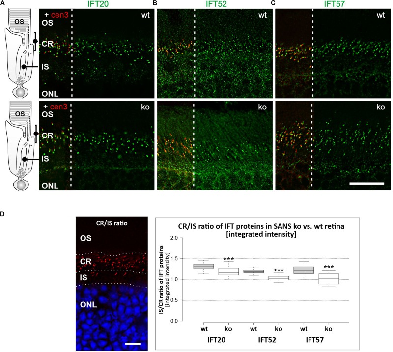
FIGURE 5.
Analysis of IFT-B protein expression in photoreceptor cells of SANS knock out (ko) mice. Immunofluorescence labelling of the photoreceptor layer of wild type mice (upper panel) and SANS ko mice (lower panel) by anti-IFT-B molecules, IFT20 (A), IFT52 (B), and IFT57 (C), respectively, counterstained for nuclear DNA by DAPI. Scale bar: 10 μm. (D) BoxPlot analysis of fluorescence intensities of anti-IFT-B staining in the ciliary region (CR) versus inner segment region (IS) in wild type and SANS ko mice. The presence of all three IFT-B molecules in the ciliary region is significantly lower in SANS ko mouse photoreceptor cells.

