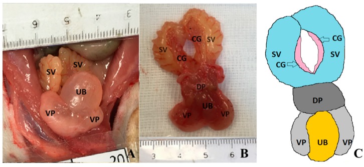
Figure 1.
Macroscopic appearance of rat prostate and other surrounding anatomical structures at 61 weeks of age. (A) In situ photography of an animal from control group, ventral view; (B) Photography of prostate from an induced animal (dorsal view). Seminal vesicles and coagulative glands extended caudally. (C) Line diagram of prostate, urinary bladder and closed sex glands. Coagulating glands (CG), dorsal (DP) and ventral prostate lobes (VP), seminal vesicles (SV) and urinary bladder (UB).

