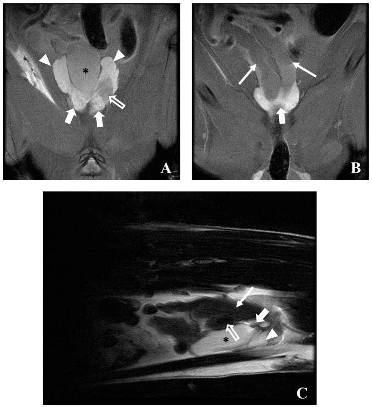
Figure 9.
MRI PD weighted images of a cancer-induced animal, 58 weeks of age in dorsal (A,B) and sagittal (C) plane. The urinary bladder (asterisk) is well visible, surrounded by the dorsal (bold arrow) and ventral prostate (arrowhead), and the seminal vesicles (arrow). Low-intensity MRI signals were observed (open arrows) in transition dorsal/ventral prostate (B) and in seminal vesicles (C).

