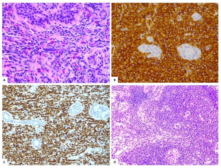
Figure 1.
(A) Isolated myeloid sarcoma (MS) involving the right breast (40×, H&E): Diffuse infiltration by blasts with occasional eosinophils. (B) The neoplastic cells show immunoreactivity for CD34 (40×). (C) Anti-TdT immunohistochemical staining (20×). (D) Fine-needle biopsy confirmed the presence of a monotonous population of blasts diagnosis of MS involving the left breast (20×, H&E).

