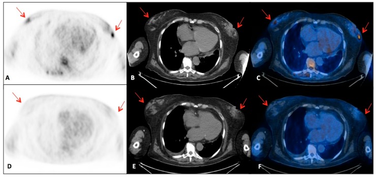
Figure 2.
18F-FDG-PET/CT examinations performed in a patient affected with breast MS: axial (A,D) PET, (B,E) CT and (C,F) fusion images. (A–C) Baseline 18F-FDG-PET/CT shows bilateral multi-focal breast lesions with increased 18F-FDG uptake (SUV max 5.4) (red arrows). (D–F) Post-radiotherapy 18F-FDG-PET/CT demonstrates disappeared of the metabolically active lesions previously described (red arrows) and complete response to therapy.

