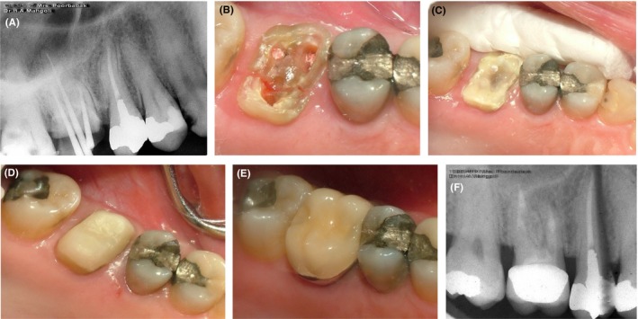
Figure 1.

Case I, A, PA radiograph of the maxillary right first molar; B, Cracks are visible in the pulpal floor after restoration removal; C, Application of Panavia cement and allowing time for cement setting (a minimum of 24 h); D, Composite build‐up and core fabrication; E, Final restoration cemented with glass ionomer cement; F, Follow‐up radiograph taken at 18 mo
