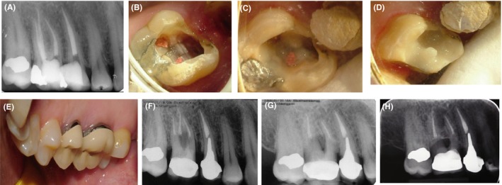
Figure 2.

Case II, A, PA radiograph of the maxillary right first molar; B, Crack lines visible in the pulp chamber floor; C, Restoring the crack lines in the pulp chamber floor with Panavia; D, Glass ionomer cement applied as base; E, Final crown fabricated for the patient; F, Final periapical radiograph; G, Follow‐up radiograph taken at 28 mo; H, Follow‐up radiograph taken at 10 y
