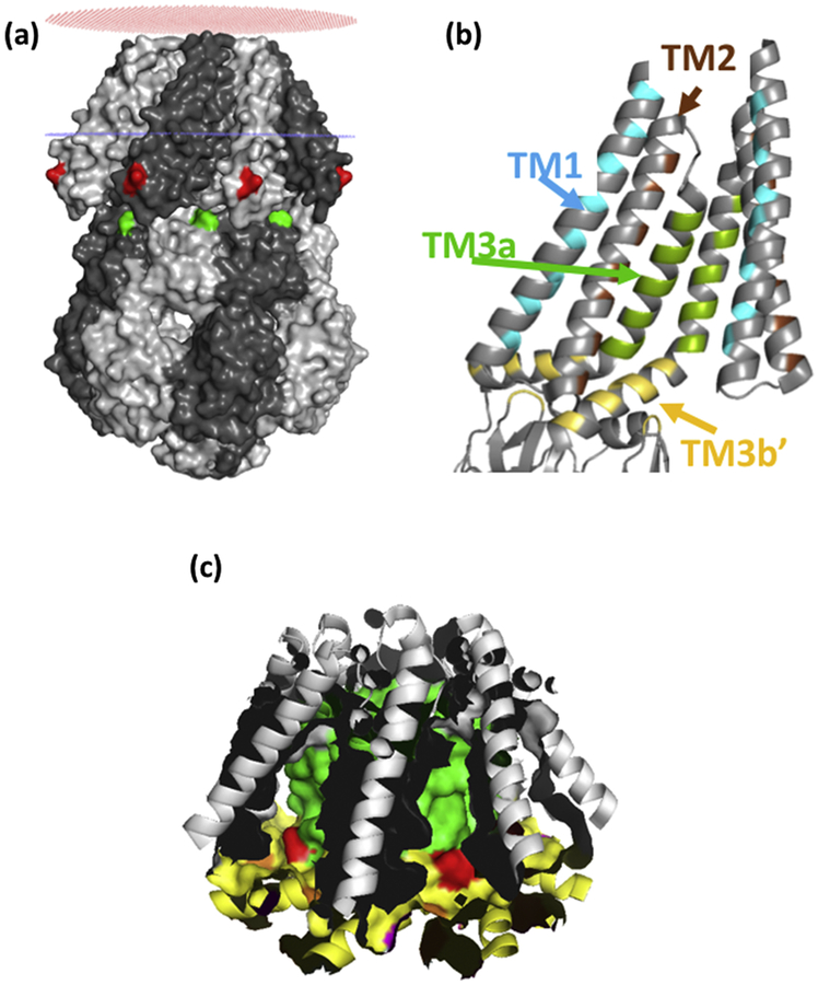
Figure 1.
(a) OPM model of surface-rendered closed structure MscS. I57 (red), and I150 (green) are marked. Red and blue rings indicate the position of the lipid/headgroup region of the periplasmic and cytoplasmic leaflets of the bilayer, respectively. (b) Hydrophobic pocket in MscS; seven identical hydrophobic pockets are created by residues on TM1 (cyan)) TM2 (brown), TM3a (green) and TM3b/beta domain (Yellow). The pocket is a volume that is contiguous with the membrane bilayer and is not a binding site for lipids. It is proposed that the lipid chains of the phospholipids that surround the periphery of MscS reversibly and dynamically penetrate the pockets. Only two subunits are shown for clarity. (c) The surface of the pockets are shown with the same colour scheme as in (b). L111 is marked in red. All images are based on the closed structure (pdb: 2OAU).

