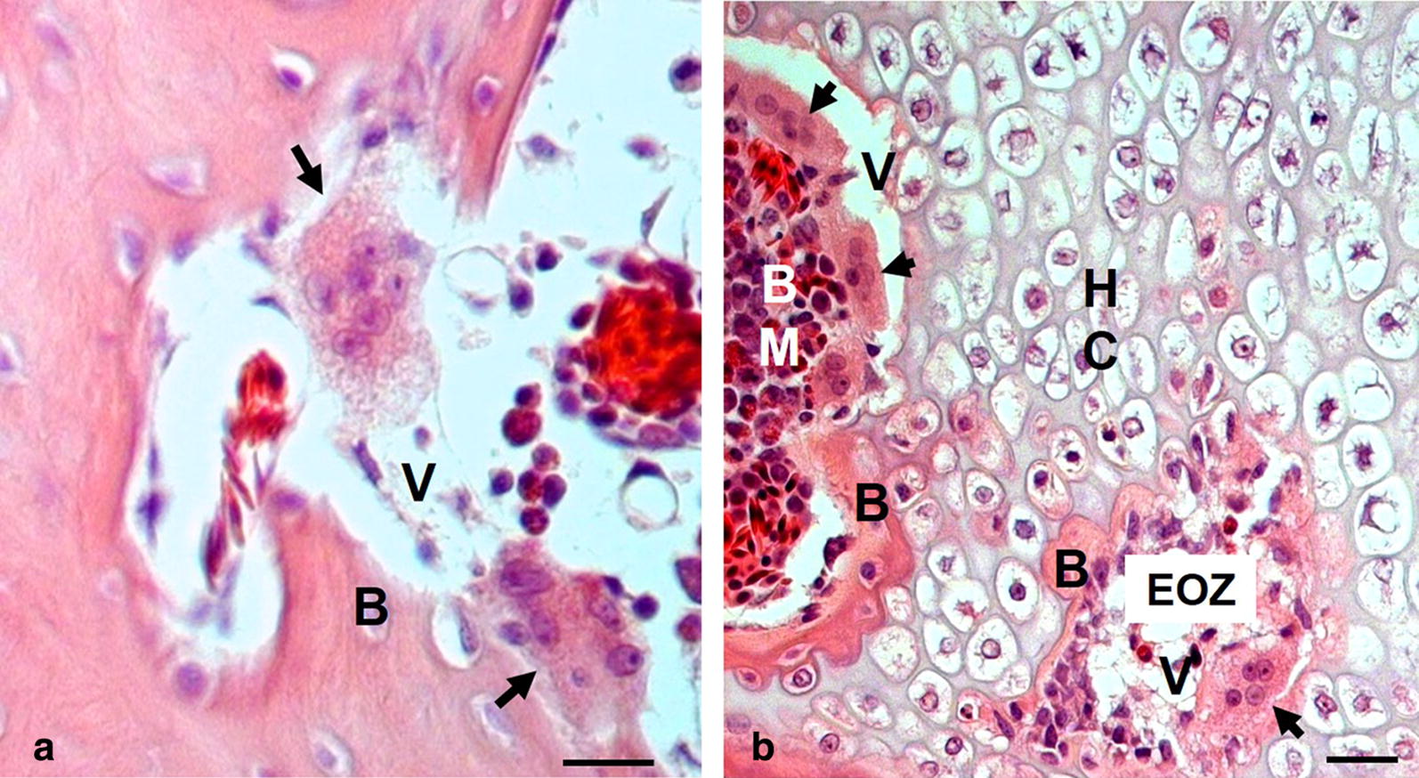
Fig. 6.

Microscopic images of cervical vertebrae sections from a broiler chicken with clinical signs of cervical spine deformity. a Erosions in newly formed bone structure associated with aggressive action of pathologically altered osteoclasts (arrows). Noteworthy are extensive amorphous vacuoles (V) in the areas previously occupied by bone structure (B). b cervical vertebrae hypertrophic cartilage (HC) with osteolysis of newly formed bone structure (B) at the interface of calcification zones of cartilage and bone marrow cavity (BM), as well as newly formed bone in the areas of endochondral ossification zone (EOZ) associated with aggressive action of pathologically altered osteoclasts, which are considerably enlarged and contain numerous nuclei (arrows). Noteworthy are clusters of osteoclasts lining the interface of bone marrow cavity and calcification zone of cartilage and penetrating the endochondral bone (arrows). It is apparent that the action of osteoclasts prevented bone ossification leaving amorphous vacuoles (V) in the areas that should be filled by newly formed bone structure (B). Bars: 20 µm
