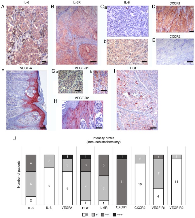
Figure 5.
Characteristic samples of expression of selected protein markers in sections from primary MM in the same cohort of patients. Multiple markers were detected by immunohistochemistry in melanomas. (A) IL-6 and (B) IL-6 receptor were detected in the majority of tumours in MM cells. (C) IL-8 expression was highly variable in studied samples; representative sections are included. Differences in expression were also observed in CXCR1 and CXCR2. (D) CXCR1 with highly positive in all patients, which contrasted with (E) the low to negative CXCR2 expression. (F) Notable positivity for VEGF was observed in the epidermis overlaying the MM lesion. Expression of VEGF receptors (G) VEGF-R1 and (H) VEGF-R2 was also observed beside MM cells in the tumour microenvironment, represented by stromal macrophages and epidermis. (I) HGF was also observed in MM cells and macrophages. Scale bar, 50 µm. (J) A summary of the expression of selected markers in primary MM in the present cohort. Semi-quantitative analysis (0, +, ++, and +++) of the immunohistochemistry reaction was used to express the proportion of positive staining based on inspection under an optical microscope. The numbers of positive tumours are included on the graph. IL, interleukin; CXCR1, C-X-C motif chemokine receptor 1; HGF, hepatocyte growth factor; CXCR2, C-X-C motif chemokine receptor 2; VEGF, vascular endothelial growth factor; VEGF-AR1, VEGF-A receptor 1; VEGF-AR2, VEGF-A receptor 2; IL-6R, IL-6 receptor.

