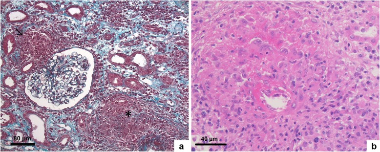
Fig. 2.
Kidney biopsy showing destruction of arteries by epithelioid cell granulomas (Masson’s trichrome (a) and H&E staining (b)). (a-b) Light microscopy. (a) Juxtaglomerular (↘) and periarteriolar (*) non caseating granulomas. Glomeruli were unremarkable (Masson trichrome staining; original magnification, × 200). (b) Segmental necrotizing granulomatous arteritis (H&E staining; original magnification, × 400)

