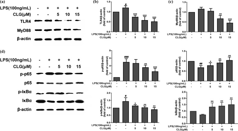
Figure 3.
CLG inhibited TLR4/NF-κB protein expression in LPS-induced BV2 microglia. BV2 cells were pretreated with CLG for 1 h and stimulated with LPS. After stimulation for 24 h, cell extracts were prepared and subjected to western blotting with TLR4 and MyD88 antibodies. After stimulation for 1 h, cell extracts were prepared and subjected to western blotting with IκBα, phospho-IκBα, NF-κB p65, and phospho-NF-κB p65 antibodies. β-Actin was used as the internal control for normalization. (a) Western blot bands of TLR4 and MyD88. (b) The density of TLR4 bands was measured, and their ratio was calculated. (c) Density ratio of MyD88 bands. (d) Western blot bands of IκBα, phospho-IκBα, NF-κB p65, and phospho-NF-κB p65. (e–h) Density ratio of phospho-NF-κB p65, p65, phospho-IκBα, and IκBα. (The results are presented as the mean ± SD from at least three independent experiments. *p < 0.05, **p < 0.01, and ***p < 0.001 compared with cells treated with LPS; #p < 0.05, ##p < 0.01, and ###p < 0.001 compared with the control.)

