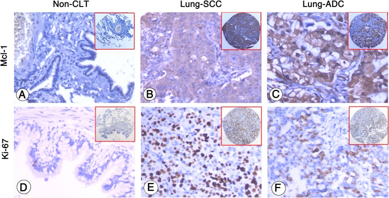
Fig. 1.
Mcl-1 expression and Ki-67 PI in non-cancerous lung tissues (non-CLT), lung SCC cells and lung ADC cells were detected by IHC using specific antibody as described in the section of materials and methods. Negative staining of Mcl-1 and low PI were showed in non-CLT (a and d, 200×, IHC, DAB staining). Strong positive staining of Mcl-1 was found in cell cytoplasm of lung SCC and lung ADC cells (b and c, 200×, IHC, DAB staining), and high PI was found in the nucleus of lung SCC and lung ADC cells (e and f, 200×, IHC, DAB staining)

