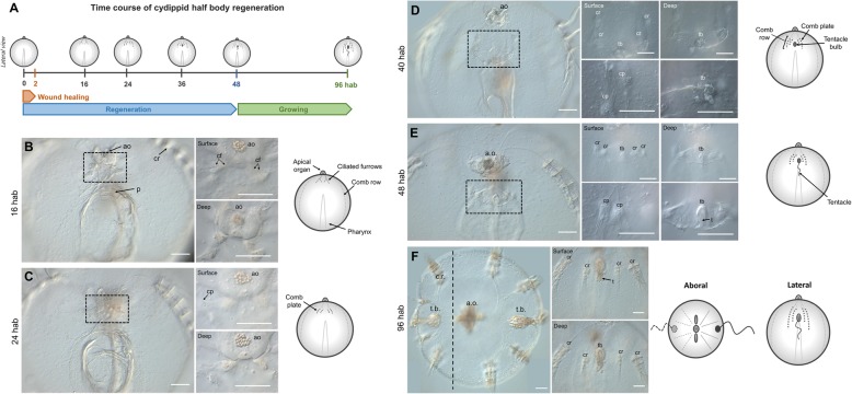
Fig. 3.
Half-body regeneration in Mnemiopsis leidyi cydippid. a Schematic representation of the time course of morphogenic events during cydippid half-body regeneration. All cartoons correspond to lateral views of the cydippid’s body bisected through the oral-aboral axis showing the cut site in the first plane. The apical organ is located at the aboral end (top) and the mouth at the oral end (down). For simplicity, tissues on the opposite body site are not depicted. b–f DIC images showing the cut site of regenerating cydippids from 16 to 96 hab. Dotted line rectangles in b–e show the area corresponding to higher magnifications on the right. Magnifications show surface and deep planes. The vertical dotted line in f indicates the approximate position of bisection, and all tissue in the left of the line is regenerated tissue. N > 100. Scale bars = 100 μm. Abbreviations: hab hours after bisection, ao apical organ, p pharynx, cr comb row, cf ciliated furrow, cp comb plate, tb tentacle bulb, t tentacle

