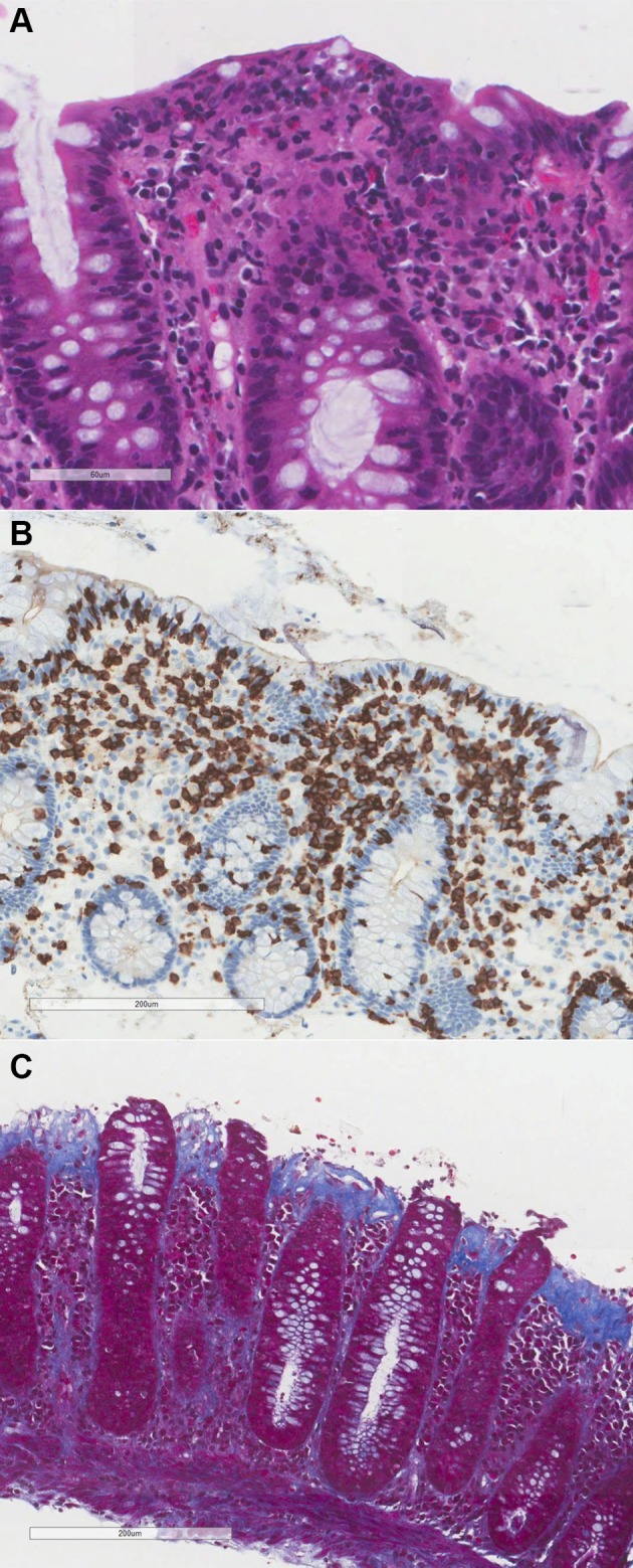
Figure 2.

Characteristic histology of lymphocytic colitis and collagenous colitis. (A) Lymphocytic colitis with increased intraepithelial lymphocytes (H&E). (B) CD3 immunohistochemistry demonstrates the increased intraepithelial lymphocytes, stained brown. CD3 antigen is specific to T-lymphocytes. (C) Thickened collagen band and loss of surface epithelium in collagenous colitis (Masson’s trichrome). Masson’s trichrome staining protocol stains the subepithelial collagen band blue.
