Figure 5.
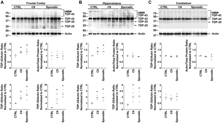
C9orf72 and sporadic ALS/FTLD have increased TDP-43 protein levels in the frontal cortex and hippocampus. Equal amounts of total proteins from the indicated human patient frontal cortex (A), hippocampus (B) and cerebellum (C) were subjected to immunoblot analysis with anti-C-terminal TDP-43 antibody and anti-actin antibodies. The protein levels of full-length TDP-43 and the C-terminal fragments TDP-35 and TDP-25 were quantified and normalized to actin, and the protein levels of actin were quantified and normalized to total protein. The mean for each experimental group was represented by a horizontal line. Quantification of TDP-25 protein levels in the cerebellum was not provided due to absence of this species in the immunoblot. *P < 0.05 compared with control based on one-way ANOVA and post-hoc Tukey’s test. C9, C9orf72; CTRL, control; HMW TDP-43, higher molecular weight species identified by the C-terminal anti-TDP-43 antibody.

