Figure 1.

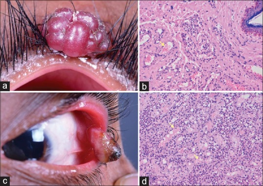
Eyelid and conjunctival lobular capillary hemangioma, (a) lobular capillary hemangioma arising from the upper eyelid of an adult male, who underwent excisional biopsy, (b) histopathology of excised specimen showing small caliber proliferating blood vessels (yellow arrow) filled with blood and adjacent stroma showing inflammatory cells, (c) lobular capillary hemangioma arising from the conjunctiva of an adult female, who underwent excisional biopsy, (d) histopathology of excised specimen showing small caliber proliferating blood vessels (yellow arrow) filled with blood separated by fibrocollagenous tissue
