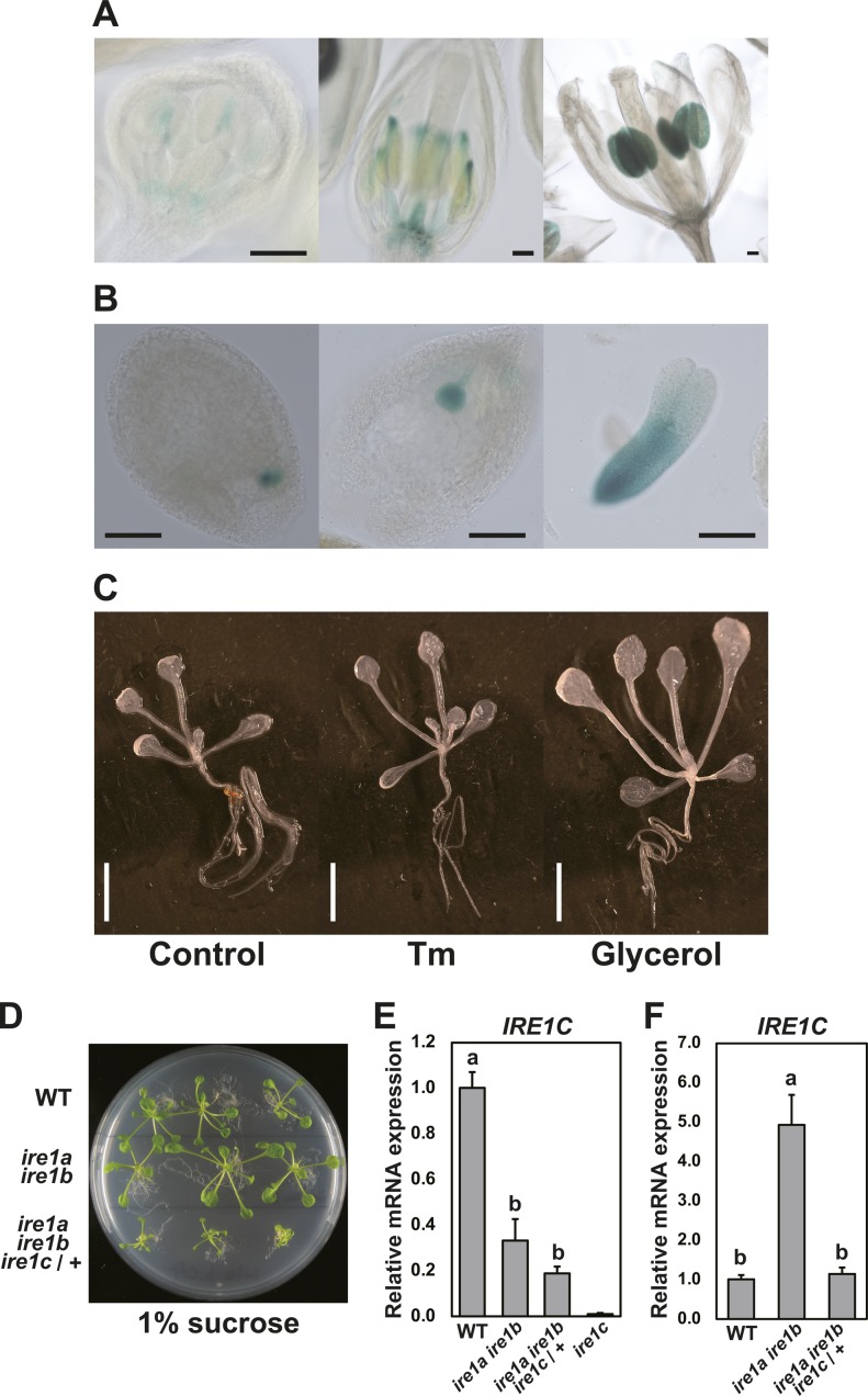
Figure S3. Tissue-specific expression of IRE1C gene.
(A, B, C) GUS histochemical staining of transgenic Arabidopsis containing IRE1C promoter–GUS fusion construct in floral tissues (A), ovules, and embryo (B). Bar = 100 μm. (C) 8-d-old seedlings treated with or without Tm for 5 h, and 11-d-old seedlings treated with glycerol for 3 d. Bar = 5 mm. (D) WT (upper), ire1a ire1b (middle), and ire1a ire1b ire1c/+ (lower) plants at 30 DAG cultured on 1/2 MS plate containing 1% sucrose. Plants were cultured on medium containing 2% sucrose for 10 d and then transferred to the 1% sucrose medium. (E, F) The relative mRNA level of IRE1C in WT, ire1a ire1b, ire1a ire1b ire1c/+, and ire1c plants (E) as indicated in (D) or flower bud tissues (F). Data are means ± SEM of four independent experiments. Different letters within each treatment indicate significant differences (P < 0.05) by the Tukey–Kramer HSD test.

