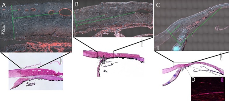
Figure 7.
Representative, tiled sections from feline anterior segment tissues IF labeled for vascular endothelial cells with vWF (red) and DAPI nuclear counterstain (blue) include differential interference contrast (DIC) signal for visualization of scleral tissue (A–C). (A) Normal eye, (B) moderately affected eye, and (C, D) severely affected eye, in which collapsed scleral vessel lumens were observed with IF (D) that were not visible on OCT. *Green line on left of each image is a line drawn perpendicularly to sclera corresponding to the termination of Descemet's membrane and two perpendicular green lines depict distance measured to first small scleral vessel lumen and first large vessel lumen.

