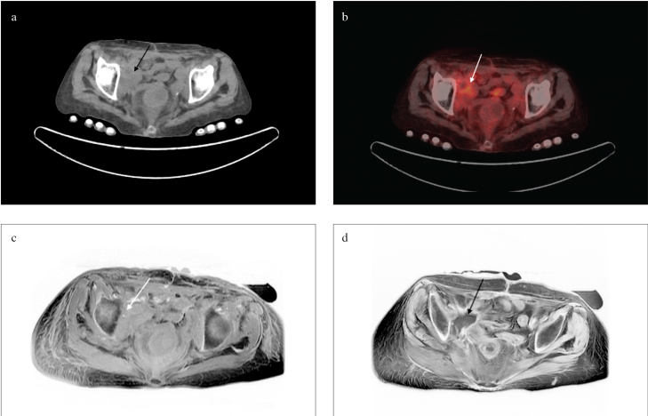
Figure 2. a–d.
Soft tissue mass (white and black arrows) adjacent to the iliac bone in the concurrent CT (a), PET/CT (b), T1 (c), and T2 (d) sequences of pelvic MRI images following cystectomy
CT: computed tomography; PET/CT: positron emission tomography/computed tomography; MRI: magnetic resonance imaging

