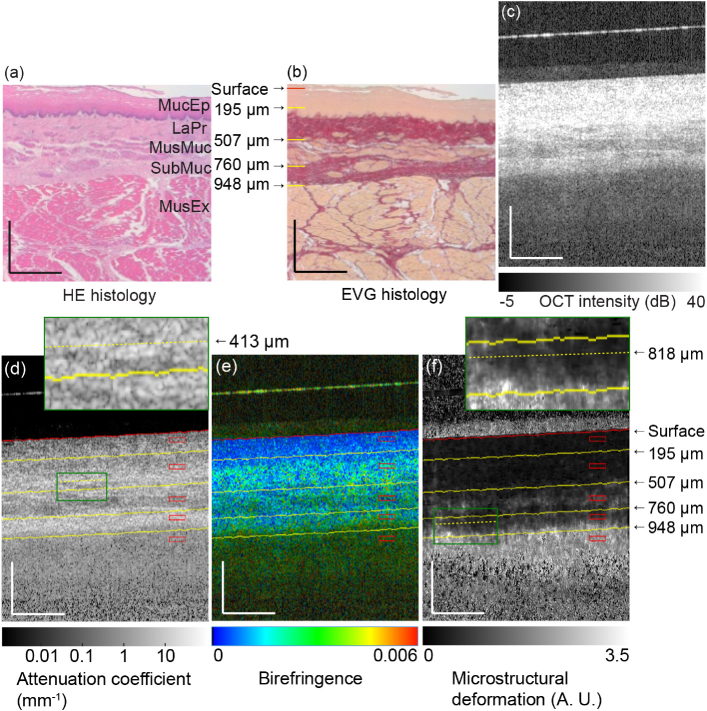
Fig. 5.
(a) H&E and (b) EVG histologies of esophagus. (c) OCT intensity, (d) attenuation coefficient, (e) birefringence, and (f) MSD images of esophagus. The scale bars represent 0.5 mm × 0.5 mm. MucEp: mucosal epithelium; LaPr: lamina propria; MusMuc: muscularis mucosa; SubMuc: submucosa; MusEx: muscularis externa. The surface of the glass plate appears as a hyperreflective line above the tissue surface in (c), (d), (e) and (f).

