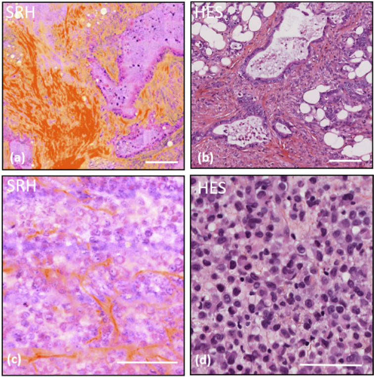
Fig. 4.
SRH/HES comparison for critical pathological situations. (a) SRH image of a cancerous omemtum, (b) standard HES of cancerous omemtum from the same patient, (c) SRH image from a thick biopsy sample, imaged directly after its gastric endoscopic excision, of a poorly differentiated adenoma case, (d) standard HES from the same patient as in (c). Scale bar is 100 µm.

