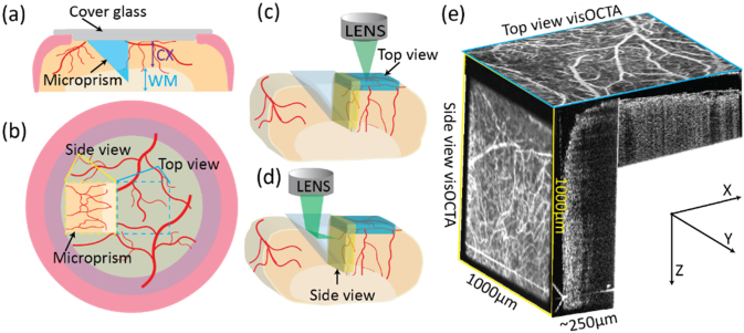
Fig. 2.
(a) Schematic of the cranial window-microprism assembly and implantation. CX: cortex; WM: white matter; (b) Relationship between the top-view and side-view images. Yellow dashed square: side-view from the microprism; blue dashed square: top-view; (c) Imaging volume acquired from top-view (blue cuboid); (d) Imaging volume acquired from side-view (yellow cuboid); (e) Illustration of top-view and side-view en face vis-OCTA images and B-scan image with respect to their geometries.

