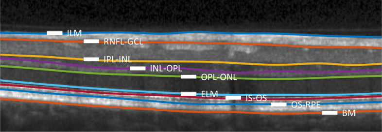
Fig. 6.
An example B-scan with manually delineated boundaries separating the following retinal layers: the retinal nerve fiber layer (RNFL), the ganglion cell layer (GCL); the inner plexiform layer (IPL); the inner nuclear layer (INL); the outer plexiform layer (OPL); the outer nuclear layer (ONL), the inner segment (IS); the outer segment (OS); and the retinal pigment epithelium (RPE). Boundaries (surfaces) between these layers are identified by hyphenating their acronyms. The named boundaries are: the inner limiting membrane (ILM); the external limiting membrane (ELM); and Bruch’s Membrane (BM).

