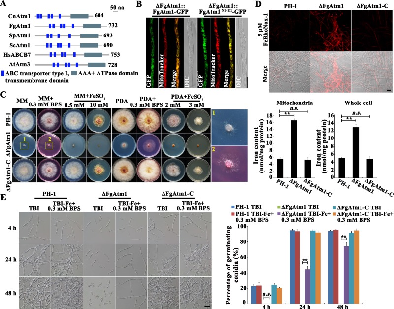
Fig 1. Deletion of FgAtm1 led to iron accumulation.
(A) Domain analyses of Atm1 orthologs from F. graminearum and other eukaryotic species. The domain analysis was performed with InterPro Scan program of the Interpro protein database (http://www.ebi.ac.uk/interpro). (B) Colocalization of FgAtm1 or FgAtm1N1-111 (truncated FgAtm1 containing only the N-terminal 111 amino acids) with mitochondrial dye MitoTracker. The plasmid FgAtm1- or FgAtm1N1-111-GFP was ectopically transformed into ΔFgAtm1, and the resulting strain was then examined with a fluorescent microscope after MitoTracker staining. Bar = 10 μm. (C) Sensitivity of the wild-type strain PH-1, ΔFgAtm1 and the complemented transformant ΔFgAtm1-C to iron chelating agent BPS and FeSO4. A 5-mm mycelial plug of each strain was inoculated on MM or PDA without or with 0.3 mM BPS or FeSO4 at the indicated concentration, and then incubated at 25°C for 3 days. (D) Iron content in mitochondria and whole cell of the wild type, ΔFgAtm1 and ΔFgAtm1-C was determined by a laser scanning microscope with 5 μΜ fluorescent iron-binding dye FeRhoNox-1 (upper panel) or colorimetric ferrozine-based assay (lower panel) after culture in CM at 25°C for 36 hours. Bar = 20 μm. Means and standard errors were calculated from three repeats. Significance was measured using unpaired t-test (n.s. not significant, **p < 0.01). (E) Conidial germination of the wild type, ΔFgAtm1 and ΔFgAtm1-C in trichothecene biosynthesis induction medium (TBI) or iron-depleted TBI. Bar = 20 μm. Percentage of germinating conidia of each strain was calculated after 4-, 24- and 48-hour-incubation. Means and standard errors were calculated from three repeats. Significance was measured using unpaired t-test (n.s. not significant, **p < 0.01).

